| Spine of scapula | |
|---|---|
 Left scapula seen from behind (spine of scapula shown in red) | |
 Position of spine of scapula shown in red | |
| Details | |
| Identifiers | |
| Latin | Spina scapulae |
| TA98 | A02.4.01.005 |
| TA2 | 1147 |
| FMA | 13453 |
| Anatomical terms of bone | |
The spine of the scapula or scapular spine is a prominent plate of bone, which crosses obliquely the medial four-fifths of the scapula at its upper part, and separates the supra- from the infraspinatous fossa.
Structure
It begins at the vertical [vertebral or medial border] border by a smooth, triangular area over which the tendon of insertion of the lower part of the Trapezius glides, and, gradually becoming more elevated, ends in the acromion, which overhangs the shoulder-joint.
The spine is triangular, and flattened from above downward, its apex being directed toward the vertebral border.
Root
The root of the spine of the scapula is the most medial part of the scapular spine. It is termed "triangular area of the spine of scapula", based on its triangular shape giving it distinguishable visible shape on x-ray images.[1] The root of the spine is on a level with the tip of the spinous process of the third thoracic vertebra.[2]
 Left scapula. Animation. Root of spine is shown in red.
 Position of root of spine (shown in red.) Animation.
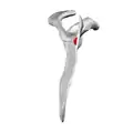 Medial view of left scapula. Root of spine shown in red.
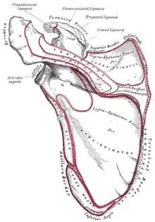 Posterior surface of scapula. Root of spine is not labeled. But visible at center right.
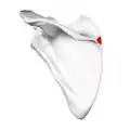 Left scapula. Posterior view. Root of spine shown in red.
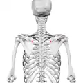 Posterior view. Root of spine shown in red.
Function
It presents two surfaces and three borders.
- Its superior surface is concave; it assists in forming the supraspinatous fossa, and gives origin to part of the supraspinatus.
- Its inferior surface forms part of the infraspinatous fossa, gives origin to a portion of the infraspinatus, and presents near its center the orifice of a nutrient canal.
Of the three borders, the anterior is attached to the dorsal surface of the bone; the posterior, or crest of the spine, is broad, and presents two lips and an intervening rough interval.
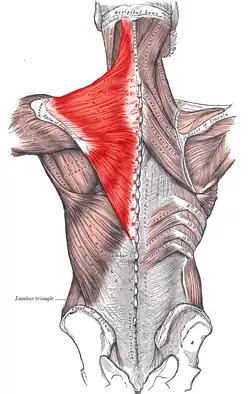
- The trapezius is attached to the superior lip, and a rough tubercle is generally seen on that portion of the spine which receives the tendon of insertion of the lower part of this muscle.
- The deltoideus is attached to the whole length of the inferior lip.
- The interval between the lips is subcutaneous and partly covered by the tendinous fibers of these muscles.
The lateral border, or base, the shortest of the three, is slightly concave; its edge, thick and round, is continuous above with the under surface of the acromion, below with the neck of the scapula. It forms the medial boundary of the great scapular notch, which serves to connect the supra- and infraspinatous fossae.
Additional image
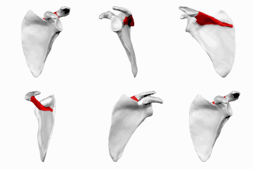 Left scapula seen from behind (spine shown in red).
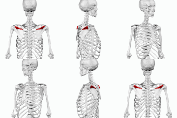 Position of spine (shown in red). Animation.
 Left scapula seen from behind (spine labeled at center top, projecting "out").
 Left scapula seen from behind (spine labeled at center top).
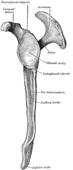
 Left scapula. Lateral view (spine shown in red)
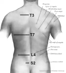 Surface anatomy of back
 Spine of scapula labeled in red, showing muscles attached to it

References
![]() This article incorporates text in the public domain from page 203 of the 20th edition of Gray's Anatomy (1918)
This article incorporates text in the public domain from page 203 of the 20th edition of Gray's Anatomy (1918)
- ↑ Al-Redouan, Azzat; Kachlik, David (2022). "Scapula revisited: new features identified and denoted by terms using consensus method of Delphi and taxonomy panel to be implemented in radiologic and surgical practice". J Shoulder Elbow Surg. 31 (2): e68-e81. doi:10.1016/j.jse.2021.07.020. PMID 34454038.
- ↑ Gray's Anatomy (1918), p.1306
External links
- Anatomy photo:03:os-0106 at the SUNY Downstate Medical Center - "Scapular Region: Scapula (Left)"