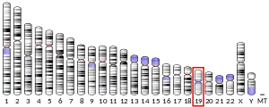
Anti-Müllerian hormone (AMH), also known as Müllerian-inhibiting hormone (MIH), is a glycoprotein hormone structurally related to inhibin and activin from the transforming growth factor beta superfamily, whose key roles are in growth differentiation and folliculogenesis.[5] In humans, it is encoded by the AMH gene, on chromosome 19p13.3,[6] while its receptor is encoded by the AMHR2 gene on chromosome 12.[7]
AMH is activated by SOX9 in the Sertoli cells of the male fetus.[8] Its expression inhibits the development of the female reproductive tract, or Müllerian ducts (paramesonephric ducts), in the male embryo, thereby arresting the development of fallopian tubes, uterus, and upper vagina.[9][10][8] AMH expression is critical to sex differentiation at a specific time during fetal development, and appears to be tightly regulated by nuclear receptor SF-1, transcription GATA factors, sex-reversal gene DAX1, and follicle-stimulating hormone (FSH).[11][12][13] Mutations in both the AMH gene and the type II AMH receptor have been shown to cause the persistence of Müllerian derivatives in males that are otherwise normally masculinized.[14]
AMH is also a product of granulosa cells of the preantral and small antral follicles in women. As such, AMH is only present in the ovary until menopause.[15] So AMH makes it possible to predict the age at which menopause will occur.Production of AMH regulates folliculogenesis by inhibiting recruitment of follicles from the resting pool in order to select for the dominant follicle, after which the production of AMH diminishes.[15][16] As a product of the granulosa cells, which envelop each egg and provide them energy, AMH can also serve as a molecular biomarker for relative size of the ovarian reserve.[17] In humans, this is helpful because the number of cells in the follicular reserve can be used to predict timing of menopause.[18] In bovine, AMH can be used for selection of females in multi-ovulatory embryo transfer programs by predicting the number of antral follicles developed to ovulation.[19] AMH can also be used as a marker for ovarian dysfunction, such as in women with polycystic ovary syndrome (PCOS).
Structure

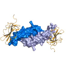
AMH is a dimeric glycoprotein with a molar mass of 140 kDa.[20] The molecule consists of two identical subunits linked by sulfide bridges, and characterized by the N-terminal dimer (pro-region) and C-terminal dimer.[5] AMH binds to its Type 2 receptor AMHR2, which phosphorylates a type I receptor under the TGF beta signaling pathway.[5]
Function
Embryogenesis
In male mammals, AMH prevents the development of the Müllerian ducts into the uterus and other Müllerian structures.[9] The effect is ipsilateral, that is each testis suppresses Müllerian development only on its own side.[21] In humans, this action takes place during the first eight weeks of gestation. If no hormone is produced from the gonads, the Müllerian will develop thanks to the presence of Wnt4 , while the Wolffian ducts, which are responsible for male reproductive parts, will die due to the presence of COUP-TFII.[22] Amounts of AMH that are measurable in the blood vary by age and sex. AMH works by interacting with specific receptors on the surfaces of the cells of target tissues (anti-Müllerian hormone receptors). The best-known and most specific effect, mediated through the AMH type II receptors, includes programmed cell death (apoptosis) of the target tissue (the fetal Müllerian ducts).
Ovarian
AMH is produced by granulosa cells from pre-antral and antral follicles, restricting expression to growing follicles, until they have reached the size and differentiation state at which they are selected for dominance by the action of pituitary FSH. Ovarian AMH expression has been observed as early as 36 weeks' gestation in the humans' fetus.[23] AMH expression is greatest in the recruitment stage of folliculogenesis, in the preantral and small antral follicles. This expression diminishes as follicles develop and enter selection stage, upon which FSH expression increases.[24] Some authorities suggest it is a measure of certain aspects of ovarian function,[25] useful in assessing conditions such as polycystic ovary syndrome and premature ovarian failure.[26]
Other
AMH production by the Sertoli cells of the testes remains high throughout childhood in males but declines to low levels during puberty and adult life. AMH has been shown to regulate production of sex hormones,[27] and changing AMH levels (rising in females, falling in males) may be involved in the onset of puberty in both sexes. Functional AMH receptors have also been found to be expressed in neurons in the brains of embryonic mice, and are thought to play a role in sexually dimorphic brain development and consequent development of gender-specific behaviours.[28] In a clade of Sebastes rockfishes in the Northwest Pacific Ocean, a duplicated copy of the AMH gene (called AMHY) is the master sex-determining gene.[29] In vitro experiments demonstrate that the overexpression of AMHY causes female-to-male sex reversal in at least one species, S. schlegelii.[29]
Pathology
In males, inadequate embryonal AMH activity can lead to persistent Müllerian duct syndrome (PMDS), in which a rudimentary uterus is present and testes are usually undescended. The AMH gene (AMH) or the gene for its receptor (AMH-RII) are usually abnormal. AMH measurements have also become widely used in the evaluation of testicular presence and function in infants with intersex conditions, ambiguous genitalia, and cryptorchidism.[30]
A study published in Nature Medicine found a link between hormonal imbalance in the womb and polycystic ovary syndrome (PCOS), specifically prenatal exposure to anti-Müllerian hormone.[31] For the study, the researchers injected pregnant mice with AMH so that they had a higher than normal concentration of the hormone. Indeed, they gave birth to daughters who later developed PCOS-like tendencies. These included problems with fertility, delayed puberty, and erratic ovulation. To reverse it, the researchers dosed the polycystic mice with an IVF drug called cetrorelix, which made the symptoms to go away. These experiments should be confirmed in humans, but it could be the first step in understanding the relationship between the polycystic ovary and the anti-Müllerian hormone.
Blood levels
In healthy females AMH is either just detectable or undetectable in cord blood at birth and demonstrates a marked rise by three months of age; while still detectable it falls until four years of age before rising linearly until eight years of age remaining fairly constant from mid-childhood to early adulthood – it does not change significantly during puberty.[32] The rise during childhood and adolescence is likely reflective of different stages of follicle development.[33] From 25 years of age AMH declines to undetectable levels at menopause.[32]
The standard measurement of AMH follows the Generation II assay. This should give the same values as the previously used IBC assay, but AMH values from the previously used DSL assay should be multiplied with 1.39 to conform to current standards because it used different antibodies.[34]
Weak evidence suggests that AMH should be measured only in the early follicular phase because of variation over the menstrual cycle. Also, AMH levels decrease under current use of oral contraceptives and current tobacco smoking.[35]
Reference ranges
Reference ranges for anti-Müllerian hormone, as estimated from reference groups in the United States, are as follows:[36]
Females:
| Age | Unit | Value |
|---|---|---|
| Younger than 24 months | ng/mL | Less than 5 |
| pmol/L | Less than 35 | |
| 24 months to 12 years | ng/mL | Less than 10 |
| pmol/L | Less than 70 | |
| 13–45 years | ng/mL | 1 to 10 |
| pmol/L | 7 to 70 | |
| More than 45 years | ng/mL | Less than 1 |
| pmol/L | Less than 7 |
Males:
| Age | Unit | Value |
|---|---|---|
| Younger than 24 months | ng/mL | 15 to 500 |
| pmol/L | 100 to 3500 | |
| 24 months to 12 years | ng/mL | 7 to 240 |
| pmol/L | 50 to 1700 | |
| More than 12 years | ng/mL | 0.7 to 20 |
| pmol/L | 5 to 140 |
AMH measurements may be less accurate if the person being measured is vitamin D deficient.[37] Note that males are born with higher AMH levels than females in order to initiate sexual differentiation, and in women, AMH levels decrease over time as fertility decreases as well.[37]
Clinical usage
General fertility assessment
Comparison of an individual's AMH level with respect to average levels[32] is useful in fertility assessment, as it provides a guide to ovarian reserve. Because one's AMH level cannot be altered by any external factors, it helps identify whether a woman needs to consider either egg freezing or trying for a pregnancy sooner rather than later if their long-term future fertility is poor.[38] A higher level of anti-Müllerian hormone when tested in women in the general population has been found to have a positive correlation with natural fertility in women aged 30–44 aiming to conceive spontaneously, even after adjusting for age.[35] However, this correlation was not found in a comparable study of younger women (aged 20 to 30 years).[35]
In vitro fertilization
AMH is a predictor for ovarian response in in vitro fertilization (IVF). Measurement of AMH supports clinical decisions, but alone it is not a strong predictor of IVF success. Women with lower levels of AMH are still able to get pregnant[39] Additionally, AMH levels are used to estimate a woman's remaining egg supply.[40]
According to NICE guidelines of in vitro fertilization, an anti-Müllerian hormone level of less than or equal to 5.4 pmol/L (0.8 ng/mL) predicts a low response to gonadotrophin stimulation in IVF, while a level greater than or equal to 25.0 pmol/L (3.6 ng/mL) predicts a high response.[41] Other cut-off values found in the literature vary between 0.7 and 20 pmol/L (0.1 and 2.97 ng/mL) for low response to ovarian hyperstimulation.[34] Subsequently, higher AMH levels are associated with greater chance of live birth after IVF, even after adjusting for age.[35][42] AMH can thereby be used to rationalise the programme of ovulation induction and decisions about the number of embryos to transfer in assisted reproduction techniques to maximise pregnancy success rates whilst minimising the risk of ovarian hyperstimulation syndrome (OHSS).[43][44] AMH can predict an excessive response in ovarian hyperstimulation with a sensitivity and specificity of 82% and 76%, respectively.[45]
Measuring AMH alone may be misleading as high levels occur in conditions like polycystic ovarian syndrome and therefore AMH levels should be considered in conjunction with a transvaginal scan of the ovaries to assess antral follicle count[46] and ovarian volume.[47]
Natural remedies
Studies into treatments to improve low ovarian reserve and low AMH levels have met with some success. Current best available evidence suggests that DHEA improves ovarian function, increases pregnancy chances and, by reducing aneuploidy, lowers miscarriage rates.[48] The studies into DHEA for low AMH show that a dose of 75 mg for a period of 16 weeks should be taken. Improvement of oocyte/embryo quality with DHEA supplementation potentially suggests a new concept of ovarian aging, where ovarian environments, but not oocytes themselves, age. DHEA has positive outcomes for women with AMH levels over 0.8 ng/mL or 5.7 pmol/L[49] DHEA has no apparent effect on oocytes or ovarian environments under this range.
Studies on CoQ10 supplementation in an aged animal model delayed depletion of ovarian reserve, restored oocyte mitochondrial gene expression, and improved mitochondrial activity.[50] Authors note that to replicate the 12–16 weeks of using CoQ10 supplements on mice to achieve these results would be the equivalent to a decade in humans.[50]
Vitamin D is believed to play a role in AMH regulation. The AMH gene promoter contains a vitamin D response element that may cause vitamin D status to influence serum AMH levels. Women with levels of vitamin D of 267.8 ± 66.4 nmol/L show a 4 times better success rate with IVF procedure than those with low levels of 104.3 ± 21 nmol/L. Vitamin D deficiency should be considered when serum AMH levels are obtained for diagnosis.[37]
Women with cancer
In women with cancer, radiation therapy and chemotherapy can damage the ovarian reserve. In such cases, a pre-treatment AMH is useful in predicting the long-term post-chemotherapy loss of ovarian function, which may indicate fertility preservation strategies such as oocyte cryopreservation.[35] A post-treatment AMH is associated with decreased fertility.[33][35]
Granulosa cell tumors of the ovary secrete AMH, and AMH testing has a sensitivity ranging between 76 and 93% in diagnosing such tumors.[35] AMH is also useful in diagnosing recurrence of granulosa cell tumors.[35]
Neutering status in animals
In veterinary medicine, AMH measurements are used to determine neutering status in male and female dogs and cats. AMH levels can also be used to diagnose cases of ovarian remnant syndrome.[51]
Biomarker of polycystic ovary syndrome
Polycystic ovary syndrome (PCOS) is an endocrine disorder most commonly found in women of reproductive age that is characterized by oligo- or anovulation, hyperandrogenism, and polycystic ovaries (PCO).[52] This endocrine disorder increases AMH levels at nearly two to three times higher in women with PCOS than in normal type women. This is often attributed to the increased follicle count number characteristic of PCOS, indicating an increase in granulosa cells since they surround each individual egg.[53] However, increased AMH levels have also been attributed not just to the increased number of follicles, but also to an increased amount of AMH produced per follicle.[54] The high levels of androgens, characteristic of PCOS, also stimulate and provide feedback for increased production of AMH, as well.[24] In this way, AMH has been increasingly considered to be a tool or biomarker that can be used to diagnose or indicate PCOS.
Biomarker of Turner syndrome
Turner syndrome is the most common sex chromosome-related inherited diseases in female around the world, with the incidence of 1 in 2000 live female births.[55] One of the significant pathological features is the premature ovarian failure, leading to amenorrhea or even infertility. Follicle stimulating hormone and inhibin B were recommended to be monitored routinely by specialists to speculate the condition of ovary. Recently, anti-Müllerian hormone is advised as a more accurate biomarker for follicular development by several researchers. The biological function of anti-Müllerian hormone in ovary is to counteract the recruitment of primordial follicles triggered by FSH, reserving the follicle pool for further recruitment and ovulation. When menopause takes place, the serum concentration of anti-Müllerian hormone will be nearly undetectable amongst normal women. Thus, variations in AMH levels during childhood may theoretically predict the duration of any given girl's reproductive life span, assuming that the speed of the continuous follicle loss is comparable between individuals.[56]
Potential future usage
AMH has been synthesized. Its ability to inhibit growth of tissue derived from the Müllerian ducts has raised hopes of usefulness in the treatment of a variety of medical conditions including endometriosis, adenomyosis, and uterine cancer. Research is underway in several laboratories. If there were more standardized AMH assays, it could potentially be used as a biomarker of polycystic ovary syndrome.[57]
In mice, an increase in AMH has been shown to reduce the number of growing follicles and thus the overall size of the ovaries. This increase in AMH production reduces primary, secondary and antral follicles without reducing the number of primordial follicles suggesting a blockade of primordial follicle activation. This may provide a viable method of contraception which protects the ovarian reserve of oocytes during chemotherapy without extracting them from the body allowing the potential for natural reproduction later in life.[58]
Names
The adjective Müllerian is written either Müllerian or müllerian, depending on the governing style guide; the derived term with the prefix anti- is then anti-Müllerian, anti-müllerian, or antimüllerian. The Müllerian ducts are named after Johannes Peter Müller.[59]
A list of the names that have been used for the antimüllerian hormone is as follows. For the sake of simplicity, this list ignores some orthographic variations; for example, it gives only one row for "Müllerian-inhibiting hormone", although there are four acceptable stylings thereof (capital M or lowercase m, hyphen or space).
| Protein name styling | Protein symbol |
|---|---|
| Anti-Müllerian hormone | AMH |
| Müllerian-inhibiting factor | MIF |
| Müllerian-inhibiting hormone | MIH |
| Müllerian-inhibiting substance | MIS |
| Müllerian duct inhibitory factor | MDIF |
| Müllerian regression factor | MRF |
| Anti-paramesonephric hormone | APH |
See also
- Alfred Jost - first postulated the existence of a non-testesterone substance that suppressed Müllerian hormone
- Nathalie Josso - discovered and named AMH
- Anti-Müllerian hormone receptor
- Freemartin - involvement of anti-Müllerian hormone in cattle twins of mixed sex
- Persistent Müllerian duct syndrome (PMDS)
- Sexual differentiation
References
- 1 2 3 GRCh38: Ensembl release 89: ENSG00000104899 - Ensembl, May 2017
- 1 2 3 GRCm38: Ensembl release 89: ENSMUSG00000035262 - Ensembl, May 2017
- ↑ "Human PubMed Reference:". National Center for Biotechnology Information, U.S. National Library of Medicine.
- ↑ "Mouse PubMed Reference:". National Center for Biotechnology Information, U.S. National Library of Medicine.
- 1 2 3 Rzeszowska M, Leszcz A, Putowski L, Hałabiś M, Tkaczuk-Włach J, Kotarski J, Polak G (2016). "Anti-Müllerian hormone: structure, properties and appliance". Ginekologia Polska. 87 (9): 669–674. doi:10.5603/gp.2016.0064. PMID 27723076.
- ↑ Cate RL, Mattaliano RJ, Hession C, Tizard R, Farber NM, Cheung A, et al. (June 1986). "Isolation of the bovine and human genes for Müllerian inhibiting substance and expression of the human gene in animal cells". Cell. 45 (5): 685–698. doi:10.1016/0092-8674(86)90783-X. PMID 3754790. S2CID 32395217.
- ↑ Imbeaud S, Faure E, Lamarre I, Mattéi MG, di Clemente N, Tizard R, et al. (December 1995). "Insensitivity to anti-müllerian hormone due to a mutation in the human anti-müllerian hormone receptor". Nature Genetics. 11 (4): 382–388. doi:10.1038/ng1295-382. PMID 7493017. S2CID 28532430.
- 1 2 Taguchi O, Cunha GR, Lawrence WD, Robboy SJ (December 1984). "Timing and irreversibility of Müllerian duct inhibition in the embryonic reproductive tract of the human male". Developmental Biology. 106 (2): 394–398. doi:10.1016/0012-1606(84)90238-0. PMID 6548718.
- 1 2 Behringer RR (1994). The in vivo roles of müllerian-inhibiting substance. Current Topics in Developmental Biology. Vol. 29. pp. 171–87. doi:10.1016/S0070-2153(08)60550-5. ISBN 9780121531294. PMID 7828438.
- ↑ Rey R, Lukas-Croisier C, Lasala C, Bedecarrás P (December 2003). "AMH/MIS: what we know already about the gene, the protein and its regulation". Molecular and Cellular Endocrinology. 211 (1–2): 21–31. doi:10.1016/j.mce.2003.09.007. PMID 14656472. S2CID 42292318.
- ↑ Shen WH, Moore CC, Ikeda Y, Parker KL, Ingraham HA (June 1994). "Nuclear receptor steroidogenic factor 1 regulates the müllerian inhibiting substance gene: a link to the sex determination cascade". Cell. 77 (5): 651–661. doi:10.1016/0092-8674(94)90050-7. PMID 8205615. S2CID 13364008.
- ↑ Nachtigal MW, Hirokawa Y, Enyeart-VanHouten DL, Flanagan JN, Hammer GD, Ingraham HA (May 1998). "Wilms' tumor 1 and Dax-1 modulate the orphan nuclear receptor SF-1 in sex-specific gene expression". Cell. 93 (3): 445–454. doi:10.1016/s0092-8674(00)81172-1. PMID 9590178. S2CID 19015882.
- ↑ Viger RS, Mertineit C, Trasler JM, Nemer M (July 1998). "Transcription factor GATA-4 is expressed in a sexually dimorphic pattern during mouse gonadal development and is a potent activator of the Müllerian inhibiting substance promoter". Development. 125 (14): 2665–2675. doi:10.1242/dev.125.14.2665. PMID 9636081.
- ↑ Belville C, Josso N, Picard JY (December 1999). "Persistence of Müllerian derivatives in males". American Journal of Medical Genetics. 89 (4): 218–223. doi:10.1002/(sici)1096-8628(19991229)89:4<218::aid-ajmg6>3.0.co;2-e. PMID 10727997.
- 1 2 Pellatt L, Rice S, Mason HD (May 2010). "Anti-Müllerian hormone and polycystic ovary syndrome: a mountain too high?". Reproduction. 139 (5): 825–833. doi:10.1530/REP-09-0415. PMID 20207725.
- ↑ Kollmann Z, Bersinger NA, McKinnon BD, Schneider S, Mueller MD, von Wolff M (March 2015). "Anti-Müllerian hormone and progesterone levels produced by granulosa cells are higher when derived from natural cycle IVF than from conventional gonadotropin-stimulated IVF". Reproductive Biology and Endocrinology. 13: 21. doi:10.1186/s12958-015-0017-0. PMC 4379743. PMID 25889012.
- ↑ Weenen C, Laven JS, Von Bergh AR, Cranfield M, Groome NP, Visser JA, et al. (February 2004). "Anti-Müllerian hormone expression pattern in the human ovary: potential implications for initial and cyclic follicle recruitment". Molecular Human Reproduction. 10 (2): 77–83. doi:10.1093/molehr/gah015. PMID 14742691.
- ↑ van Disseldorp J, Faddy MJ, Themmen AP, de Jong FH, Peeters PH, van der Schouw YT, Broekmans FJ (June 2008). "Relationship of serum antimüllerian hormone concentration to age at menopause". The Journal of Clinical Endocrinology and Metabolism. 93 (6): 2129–2134. doi:10.1210/jc.2007-2093. PMID 18334591.
- ↑ Rico C, Médigue C, Fabre S, Jarrier P, Bontoux M, Clément F, Monniaux D (March 2011). "Regulation of anti-Müllerian hormone production in the cow: a multiscale study at endocrine, ovarian, follicular, and granulosa cell levels". Biology of Reproduction. 84 (3): 560–571. doi:10.1095/biolreprod.110.088187. PMID 21076084.
- ↑ Hampl R, Šnajderová M, Mardešić T (2011). "Antimüllerian hormone (AMH) not only a marker for prediction of ovarian reserve". Physiological Research. 60 (2): 217–223. doi:10.33549/physiolres.932076. PMID 21114374.
- ↑ Boron WF (2003). Medical Physiology: A Cellular and Molecular Approaoch. Elsevier/Saunders. p. 1114. ISBN 978-1-4160-2328-9.
- ↑ An Introduction to Behavioral Endocrinology, Randy J Nelson, 3rd edition, Sinauer
- ↑ La Marca A, Sighinolfi G, Radi D, Argento C, Baraldi E, Artenisio AC, et al. (30 September 2009). "Anti-Mullerian hormone (AMH) as a predictive marker in assisted reproductive technology (ART)". Human Reproduction Update. 16 (2): 113–130. doi:10.1093/humupd/dmp036. PMID 19793843.
- 1 2 Dumont A, Robin G, Catteau-Jonard S, Dewailly D (December 2015). "Role of Anti-Müllerian Hormone in pathophysiology, diagnosis and treatment of Polycystic Ovary Syndrome: a review". Reproductive Biology and Endocrinology. 13: 137. doi:10.1186/s12958-015-0134-9. PMC 4687350. PMID 26691645.
- ↑ Broer SL, Eijkemans MJ, Scheffer GJ, van Rooij IA, de Vet A, Themmen AP, et al. (August 2011). "Anti-mullerian hormone predicts menopause: a long-term follow-up study in normoovulatory women". The Journal of Clinical Endocrinology and Metabolism. 96 (8): 2532–2539. doi:10.1210/jc.2010-2776. PMID 21613357.
- ↑ Visser JA, de Jong FH, Laven JS, Themmen AP (January 2006). "Anti-Müllerian hormone: a new marker for ovarian function". Reproduction. 131 (1): 1–9. doi:10.1530/rep.1.00529. PMID 16388003.
- ↑ Trbovich AM, Martinelle N, O'Neill FH, Pearson EJ, Donahoe PK, Sluss PM, Teixeira J (October 2004). "Steroidogenic activities in MA-10 Leydig cells are differentially altered by cAMP and Müllerian inhibiting substance". The Journal of Steroid Biochemistry and Molecular Biology. 92 (3): 199–208. doi:10.1016/j.jsbmb.2004.07.002. PMID 15555913. S2CID 209392.
- ↑ Wang PY, Protheroe A, Clarkson AN, Imhoff F, Koishi K, McLennan IS (April 2009). "Müllerian inhibiting substance contributes to sex-linked biases in the brain and behavior". Proceedings of the National Academy of Sciences of the United States of America. 106 (17): 7203–7208. Bibcode:2009PNAS..106.7203W. doi:10.1073/pnas.0902253106. PMC 2678437. PMID 19359476.
- 1 2 Song W, Xie Y, Sun M, Li X, Fitzpatrick CK, Vaux F, et al. (July 2021). "A duplicated amh is the master sex-determining gene for Sebastes rockfish in the Northwest Pacific". Open Biology. 11 (7): 210063. doi:10.1098/rsob.210063. PMC 8277470. PMID 34255977.
- ↑ Loeff DS, Imbeaud S, Reyes HM, Meller JL, Rosenthal IM (January 1994). "Surgical and genetic aspects of persistent müllerian duct syndrome". Journal of Pediatric Surgery. 29 (1): 61–65. doi:10.1016/0022-3468(94)90525-8. PMID 7907140.
- ↑ Tata B, Mimouni NE, Barbotin AL, Malone SA, Loyens A, Pigny P, et al. (June 2018). "Elevated prenatal anti-Müllerian hormone reprograms the fetus and induces polycystic ovary syndrome in adulthood". Nature Medicine. 24 (6): 834–846. doi:10.1038/s41591-018-0035-5. PMC 6098696. PMID 29760445.
- 1 2 3 Kelsey TW, Wright P, Nelson SM, Anderson RA, Wallace WH (2011). "A validated model of serum anti-müllerian hormone from conception to menopause". PLOS ONE. 6 (7): e22024. Bibcode:2011PLoSO...622024K. doi:10.1371/journal.pone.0022024. PMC 3137624. PMID 21789206.
- 1 2 Dewailly D, Andersen CY, Balen A, Broekmans F, Dilaver N, Fanchin R, et al. (2014). "The physiology and clinical utility of anti-Mullerian hormone in women". Human Reproduction Update. 20 (3): 370–385. doi:10.1093/humupd/dmt062. hdl:10023/7488. PMID 24430863.
- 1 2 La Marca A, Sunkara SK (2013). "Individualization of controlled ovarian stimulation in IVF using ovarian reserve markers: from theory to practice". Human Reproduction Update. 20 (1): 124–140. doi:10.1093/humupd/dmt037. PMID 24077980.
- 1 2 3 4 5 6 7 8 Broer SL, Broekmans FJ, Laven JS, Fauser BC (2014). "Anti-Müllerian hormone: ovarian reserve testing and its potential clinical implications". Human Reproduction Update. 20 (5): 688–701. doi:10.1093/humupd/dmu020. PMID 24821925.
- ↑ For mass values:
- Anti-Müllerian Hormone (AMH), Serum Archived 29 July 2013 at the Wayback Machine from Mayo Medical Laboratories. Retrieved April 2012.
- Hampl R, Šnajderová M, Mardešić T (2011). "Antimüllerian hormone (AMH) not only a marker for prediction of ovarian reserve". Physiological Research. 60 (2): 217–223. doi:10.33549/physiolres.932076. PMID 21114374.
- 1 2 3 Dennis NA, Houghton LA, Jones GT, van Rij AM, Morgan K, McLennan IS (July 2012). "The level of serum anti-Müllerian hormone correlates with vitamin D status in men and women but not in boys". The Journal of Clinical Endocrinology and Metabolism. 97 (7): 2450–2455. doi:10.1210/jc.2012-1213. PMID 22508713.
- ↑ Cupisti S, Dittrich R, Mueller A, Strick R, Stiegler E, Binder H, et al. (December 2007). "Correlations between anti-müllerian hormone, inhibin B, and activin A in follicular fluid in IVF/ICSI patients for assessing the maturation and developmental potential of oocytes". European Journal of Medical Research. 12 (12): 604–608. PMID 18024272.
- ↑ Gnoth C, Schuring AN, Friol K, Tigges J, Mallmann P, Godehardt E (June 2008). "Relevance of anti-Mullerian hormone measurement in a routine IVF program". Human Reproduction. 23 (6): 1359–1365. doi:10.1093/humrep/den108. PMID 18387961.
- ↑ Indichova J. "Does a Low AMH Level (Anti-Mullerian Hormone) Indicate Infertility?". fertileheart.com. Retrieved 6 February 2015.
- ↑ Fertility: assessment and treatment for people with fertility problems. NICE clinical guideline CG156 - Issued: February 2013
- ↑ Iliodromiti S, Kelsey TW, Wu O, Anderson RA, Nelson SM (2014). "The predictive accuracy of anti-Müllerian hormone for live birth after assisted conception: a systematic review and meta-analysis of the literature". Human Reproduction Update. 20 (4): 560–570. doi:10.1093/humupd/dmu003. PMID 24532220.
- ↑ Nelson SM, Yates RW, Fleming R (September 2007). "Serum anti-Müllerian hormone and FSH: prediction of live birth and extremes of response in stimulated cycles--implications for individualization of therapy". Human Reproduction. 22 (9): 2414–2421. doi:10.1093/humrep/dem204. PMID 17636277.
- ↑ Nelson SM, Yates RW, Lyall H, Jamieson M, Traynor I, Gaudoin M, et al. (April 2009). "Anti-Müllerian hormone-based approach to controlled ovarian stimulation for assisted conception". Human Reproduction. 24 (4): 867–875. doi:10.1093/humrep/den480. PMID 19136673.
- ↑ Broer SL, Dólleman M, Opmeer BC, Fauser BC, Mol BW, Broekmans FJ (2011). "AMH and AFC as predictors of excessive response in controlled ovarian hyperstimulation: a meta-analysis". Human Reproduction Update. 17 (1): 46–54. doi:10.1093/humupd/dmq034. PMID 20667894.
- ↑ Seifer DB, Maclaughlin DT (September 2007). "Mullerian Inhibiting Substance is an ovarian growth factor of emerging clinical significance". Fertility and Sterility. 88 (3): 539–546. doi:10.1016/j.fertnstert.2007.02.014. PMID 17559842.
- ↑ Wallace WH, Kelsey TW (July 2004). "Ovarian reserve and reproductive age may be determined from measurement of ovarian volume by transvaginal sonography". Human Reproduction. 19 (7): 1612–1617. doi:10.1093/humrep/deh285. PMID 15205396.
- ↑ Gleicher N, Barad DH (May 2011). "Dehydroepiandrosterone (DHEA) supplementation in diminished ovarian reserve (DOR)". Reproductive Biology and Endocrinology. 9: 67. doi:10.1186/1477-7827-9-67. PMC 3112409. PMID 21586137.
- ↑ Gleicher N, Weghofer A, Barad DH (September 2010). "Improvement in diminished ovarian reserve after dehydroepiandrosterone supplementation". Reproductive Biomedicine Online. 21 (3): 360–365. doi:10.1016/j.rbmo.2010.04.006. PMID 20638339.
- 1 2 Ben-Meir A, Burstein E, Borrego-Alvarez A, Chong J, Wong E, Yavorska T, et al. (October 2015). "Coenzyme Q10 restores oocyte mitochondrial function and fertility during reproductive aging". Aging Cell. 14 (5): 887–895. doi:10.1111/acel.12368. PMC 4568976. PMID 26111777.
- ↑ Place NJ, Hansen BS, Cheraskin JL, Cudney SE, Flanders JA, Newmark AD, et al. (May 2011). "Measurement of serum anti-Müllerian hormone concentration in female dogs and cats before and after ovariohysterectomy". Journal of Veterinary Diagnostic Investigation. 23 (3): 524–527. doi:10.1177/1040638711403428. PMID 21908283.
- ↑ Azziz R (March 2006). "Controversy in clinical endocrinology: diagnosis of polycystic ovarian syndrome: the Rotterdam criteria are premature". The Journal of Clinical Endocrinology and Metabolism. 91 (3): 781–785. doi:10.1210/jc.2005-2153. PMID 16418211.
- ↑ Dewailly D (November 2016). "Diagnostic criteria for PCOS: Is there a need for a rethink?". Best Practice & Research. Clinical Obstetrics & Gynaecology. 37: 5–11. doi:10.1016/j.bpobgyn.2016.03.009. PMID 27151631.
- ↑ Verma AK, Rajbhar S, Mishra J, Gupta M, Sharma M, Deshmukh G, Ali W (December 2016). "Anti-Mullerian Hormone: A Marker of Ovarian Reserve and its Association with Polycystic Ovarian Syndrome". Journal of Clinical and Diagnostic Research. 10 (12): QC10–QC12. doi:10.7860/JCDR/2016/20370.8988. PMC 5296514. PMID 28208941.
- ↑ Backeljauw P. "Clinical manifestations and diagnosis of Turner syndrome". UpToDate. Wolters Kluwer. Retrieved 1 November 2019.
- ↑ Hagen CP, Aksglaede L, Sørensen K, Main KM, Boas M, Cleemann L, et al. (November 2010). "Serum levels of anti-Müllerian hormone as a marker of ovarian function in 926 healthy females from birth to adulthood and in 172 Turner syndrome patients". The Journal of Clinical Endocrinology and Metabolism. The Journal of Clinical Endocrinology&Metabolism. 95 (11): 5003–5010. doi:10.1210/jc.2010-0930. PMID 20719830.
- ↑ Dewailly D, Lujan ME, Carmina E, Cedars MI, Laven J, Norman RJ, Escobar-Morreale HF (2013). "Definition and significance of polycystic ovarian morphology: a task force report from the Androgen Excess and Polycystic Ovary Syndrome Society". Human Reproduction Update. 20 (3): 334–352. doi:10.1093/humupd/dmt061. PMID 24345633.
- ↑ Kano M, Sosulski AE, Zhang L, Saatcioglu HD, Wang D, Nagykery N, et al. (February 2017). "AMH/MIS as a contraceptive that protects the ovarian reserve during chemotherapy". Proceedings of the National Academy of Sciences of the United States of America. 114 (9): E1688–E1697. Bibcode:2017PNAS..114E1688K. doi:10.1073/pnas.1620729114. PMC 5338508. PMID 28137855.
- ↑ Minkoff E, Baker P (2004). Biology Today: An Issues Approach (Third ed.). New York: Garland Science. p. 296. ISBN 978-1-136-83875-0.


