| BAR domain | |||||||||
|---|---|---|---|---|---|---|---|---|---|
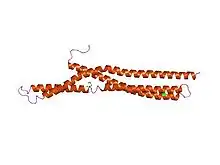
 Structure of amphiphysin BAR.[1] | |||||||||
| Identifiers | |||||||||
| Symbol | BAR | ||||||||
| Pfam | PF03114 | ||||||||
| InterPro | IPR004148 | ||||||||
| SMART | SM00721 | ||||||||
| PROSITE | PDOC51021 | ||||||||
| SCOP2 | 1uru / SCOPe / SUPFAM | ||||||||
| CDD | cd07307 | ||||||||
| |||||||||
| Bin/amphiphysin/Rvs domain | |||||||||
|---|---|---|---|---|---|---|---|---|---|
| Identifiers | |||||||||
| Symbol | BAR-2 | ||||||||
| Pfam | PF10455 | ||||||||
| |||||||||
| BAR domain of APPL family | |||||||||
|---|---|---|---|---|---|---|---|---|---|
| Identifiers | |||||||||
| Symbol | BAR-3 | ||||||||
| Pfam | PF16746 | ||||||||
| |||||||||
| EFC/F-BAR homology domain | |||||||||
|---|---|---|---|---|---|---|---|---|---|
| Identifiers | |||||||||
| Symbol | FCH | ||||||||
| Pfam | PF00611 | ||||||||
| |||||||||
| Vps5 C terminal like (BAR domain) | |||||||||
|---|---|---|---|---|---|---|---|---|---|
| Identifiers | |||||||||
| Symbol | Vps5 | ||||||||
| Pfam | PF09325 | ||||||||
| InterPro | IPR015404 | ||||||||
| |||||||||
| WASP-binding domain of sorting nexin proteins | |||||||||
|---|---|---|---|---|---|---|---|---|---|
| Identifiers | |||||||||
| Symbol | BAR-3-WASP | ||||||||
| Pfam | PF10456 | ||||||||
| |||||||||
In molecular biology, BAR domains are highly conserved protein dimerisation domains that occur in many proteins involved in membrane dynamics in a cell. The BAR domain is banana-shaped and binds to membrane via its concave face. It is capable of sensing membrane curvature by binding preferentially to curved membranes. BAR domains are named after three proteins that they are found in: Bin, Amphiphysin and Rvs.
BAR domains occur in combinations with other domains
Many BAR family proteins contain alternative lipid specificity domains that help target these protein to particular membrane compartments. Some also have SH3 domains that bind to dynamin and thus proteins like amphiphysin and endophilin are implicated in the orchestration of vesicle scission.
N-BAR domain
Some BAR domain containing proteins have an N-terminal amphipathic helix preceding the BAR domain. This helix inserts (like in the epsin ENTH domain) into the membrane and induces curvature, which is stabilised by the BAR dimer. Amphiphysin, endophilin, BRAP1/bin2 and nadrin are examples of such proteins containing an N-BAR. The Drosophila amphiphysin N-BAR (DA-N-BAR) is an example of a protein with a preference for negatively charged surfaces.[1]
Human proteins containing this domain
AMPH; ARHGAP17; ARHGAP44; BIN1; BIN2; BIN3; SH3BP1; SH3GL1; SH3GL2; SH3GL3; SH3GLB1; SH3GLB2.[2]
F-BAR (EFC) domain
F-BAR domains (for FCH-BAR, or EFC for Extended FCH Homology) are BAR domains that are extensions of the already established FCH domain. They are frequently found at the amino terminus of proteins. They can bind lipid membranes and can tubulate lipids in vitro and in vivo, but their exact physiological role still is under investigation.[3] Examples of the F-BAR domain family are CIP4/FBP17/Toca-1, Syndapins (also called PACSINs) and muniscins. Gene knock-out of syndapin I in mice revealed that this brain-enriched isoform of the syndapin family is crucial for proper size control of synaptic vesicles and thereby indeed helps to define membrane curvature a physiological process. Work of the lab of Britta Qualmann also demonstrated that syndapin I is crucial for proper targeting of the large GTPase dynamin to membranes.[4]
Sorting nexins
The sorting nexin family of proteins includes several members that possess a BAR domain, including the well characterized SNX1 and SNX9.
Human proteins containing this domain
AMPH; ARHGAP17; BIN1; BIN2; BIN3; DNMBP; GMIP; RICH2; SH3BP1; SH3GL1; SH3GL2; SH3GL3; SH3GLB1; SH3GLB2;
See also
- Arfaptin, includes a BAR-like domain
- IMD domain, a BAR-like domain
- SNX8 - protein family with a combination of BAR-like and PX domains
- Epsin
- Membrane curvature
External links
References
- 1 2 Peter BJ, Kent HM, Mills IG, et al. (January 2004). "BAR domains as sensors of membrane curvature: the amphiphysin BAR structure". Science. 303 (5657): 495–9. doi:10.1126/science.1092586. PMID 14645856. S2CID 6104655.
- ↑ "Gene group: N-BAR domain containing". HGNC: HUGO gene nomenclature committee.
- ↑ Qualmann B, Koch D, Kessels MM (August 2011). "Let's go bananas: revisiting the endocytic BAR code". EMBO J. 30 (17): 3501–15. doi:10.1038/emboj.2011.266. PMC 3181480. PMID 21878992.
- ↑ Koch D, Spiwoks-Becker I, Sabanov V, Sinning A, Dugladze T, Stellmacher A, Ahuja R, Grimm J, Schüler S, Müller A, Angenstein F, Ahmed T, Diesler A, Moser M, Tom Dieck S, Spessert R, Boeckers TM, Fässler R, Hübner CA, Balschun D, Gloveli T, Kessels MM, Qualmann B (December 2011). "Proper synaptic vesicle formation and neuronal network activity critically rely on syndapin I". EMBO J. 30 (24): 4955–69. doi:10.1038/emboj.2011.339. PMC 3243622. PMID 21926968.
Further reading
- Leventis PA, Chow BM, Stewart BA, Iyengar B, Campos AR, Boulianne GL (November 2001). "Drosophila Amphiphysin is a post-synaptic protein required for normal locomotion but not endocytosis". Traffic. 2 (11): 839–50. doi:10.1034/j.1600-0854.2001.21113.x. PMID 11733051.
- Zhang B, Zelhof AC (July 2002). "Amphiphysins: raising the BAR for synaptic vesicle recycling and membrane dynamics. Bin-Amphiphysin-Rvsp". Traffic. 3 (7): 452–60. doi:10.1034/j.1600-0854.2002.30702.x. PMID 12047553.Review.
- Zelhof AC, Bao H, Hardy RW, Razzaq A, Zhang B, Doe CQ (December 2001). "Drosophila Amphiphysin is implicated in protein localization and membrane morphogenesis but not in synaptic vesicle endocytosis". Development. 128 (24): 5005–15. PMID 11748137.
- Mathew D, Popescu A, Budnik V (November 2003). "Drosophila amphiphysin functions during synaptic Fasciclin II membrane cycling". J. Neurosci. 23 (33): 10710–6. doi:10.1523/JNEUROSCI.23-33-10710.2003. PMC 6740931. PMID 14627656.
- Peter BJ, Kent HM, Mills IG, et al. (January 2004). "BAR domains as sensors of membrane curvature: the amphiphysin BAR structure". Science. 303 (5657): 495–9. doi:10.1126/science.1092586. PMID 14645856. S2CID 6104655.
- Weissenhorn W (August 2005). "Crystal structure of the endophilin-A1 BAR domain". J. Mol. Biol. 351 (3): 653–61. doi:10.1016/j.jmb.2005.06.013. PMID 16023669.
- Gallop JL, Jao CC, Kent HM, et al. (June 2006). "Mechanism of endophilin N-BAR domain-mediated membrane curvature". EMBO J. 25 (12): 2898–910. doi:10.1038/sj.emboj.7601174. PMC 1500843. PMID 16763559.
- Masuda M, Takeda S, Sone M, et al. (June 2006). "Endophilin BAR domain drives membrane curvature by two newly identified structure-based mechanisms". EMBO J. 25 (12): 2889–97. doi:10.1038/sj.emboj.7601176. PMC 1500852. PMID 16763557.
- Frost A, Perera R, Roux A, et al. (March 2008). "Structural basis of membrane invagination by F-BAR domains". Cell. 132 (5): 807–17. doi:10.1016/j.cell.2007.12.041. PMC 2384079. PMID 18329367.